June 2, 2025 Published by Intel Newsroom

Created by Acer Medical in partnership with medical specialists, the technology is available in 13 countries.
A comprehensive eye exam in a crowded doctor’s office can often take the better part of two hours, with much of that time spent waiting for dilation-related eye drops to take effect before tests even begin.
While an initial diagnosis using fundus photography can be provided on the spot, analyzing results might take much longer: A patient may wait up to four weeks for a confirmed diagnosis, especially if the data requires input from other eye specialists.
Today, thanks to Acer Medical’s artificial intelligence-assisted diagnostic software and a combination of Intel hardware- and software-based technology, accurate results can be delivered in just seconds.
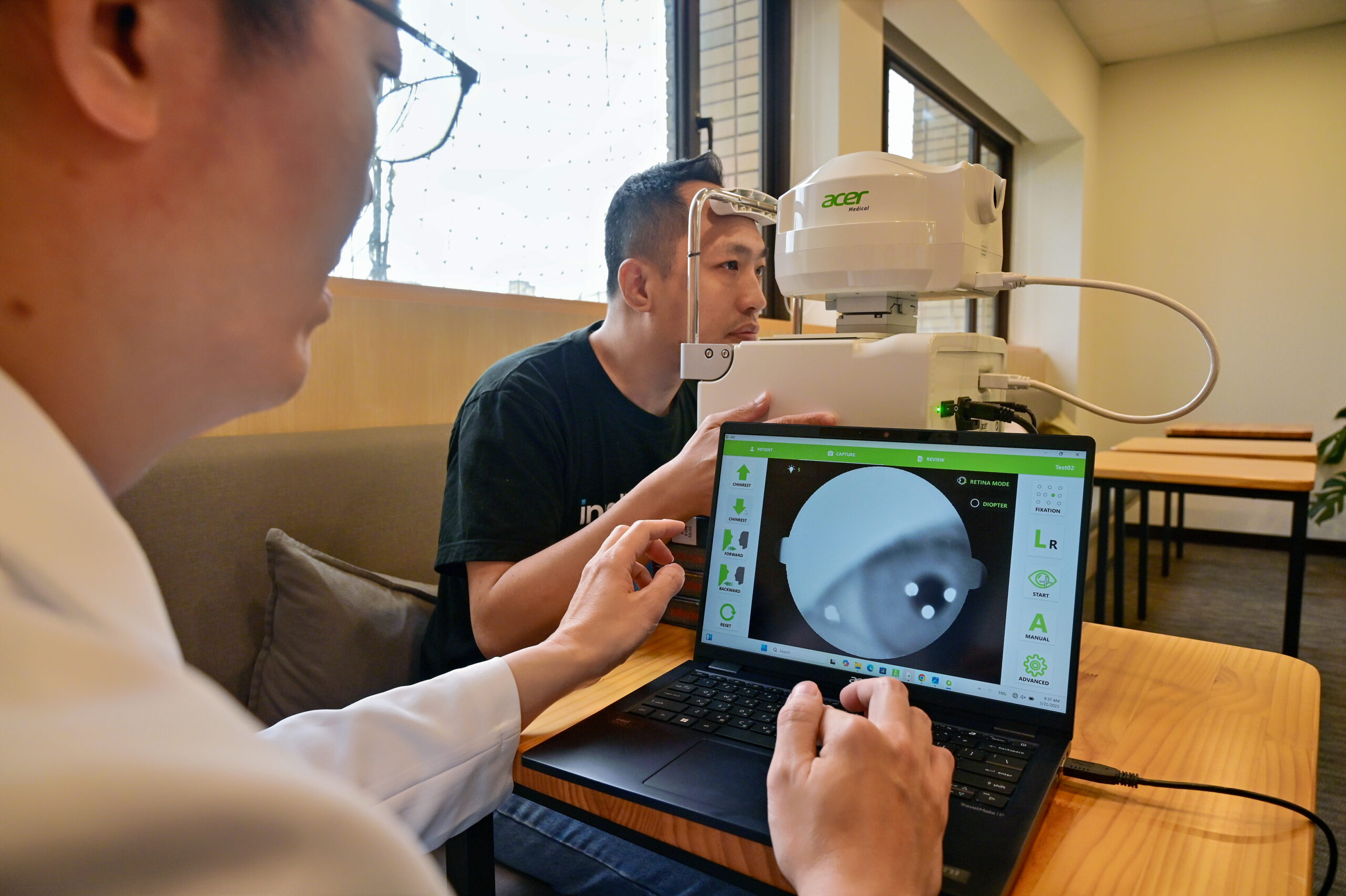
Using Acer Medical’s VeriSee DR for diabetic retinopathy and VeriSee Age-related Macular Degeneration (VeriSee AMD) for age-related macular degeneration, results are processed and stored on an Acer laptop powered by Intel® Core™ Ultra processors, with no Internet connection required.
These tools boost doctor productivity and efficiency while keeping patient data private in a secure medical environment, providing additional peace of mind. And because all the AI data-crunching happens on the AI PC, it can be performed away from a hospital setting – bringing diagnoses to people who are far from healthcare specialists.
AI-Powered Solutions Quicken Diagnoses
VeriSee DR and VeriSee AMD – technologies available today in 13 countries – provide health care professionals with new tools powered by AI-based algorithms to shorten diagnosis times and expedite patient care.
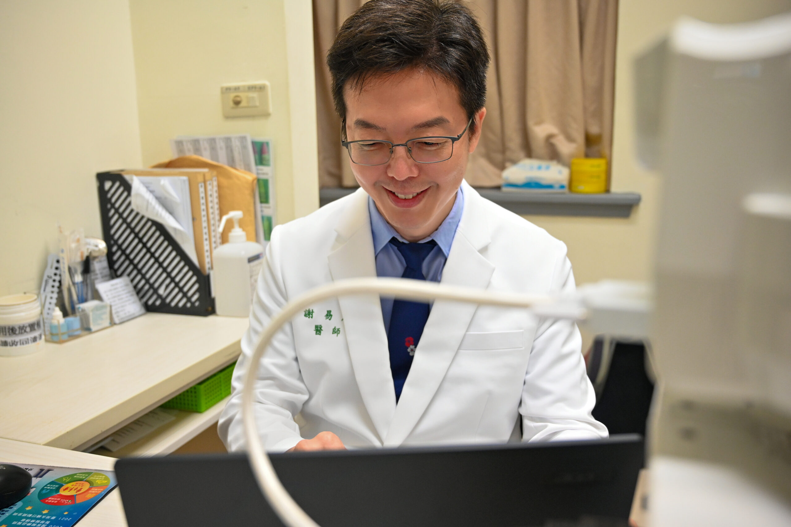
Dr. Hsieh Yi-Ting, a consultant ophthalmologist and retina specialist at the National Taiwan University Hospital, has spent more than two decades in the field. He developed VeriSee DR and advocated for its use across Taiwan to quickly assess a patient for diabetic retinopathy, an eye condition that causes vision loss and blindness in diabetic patients.
He also developed VeriSee AMD to detect age-related macular degeneration, an eye disease that progressively blurs vision as people grow older.
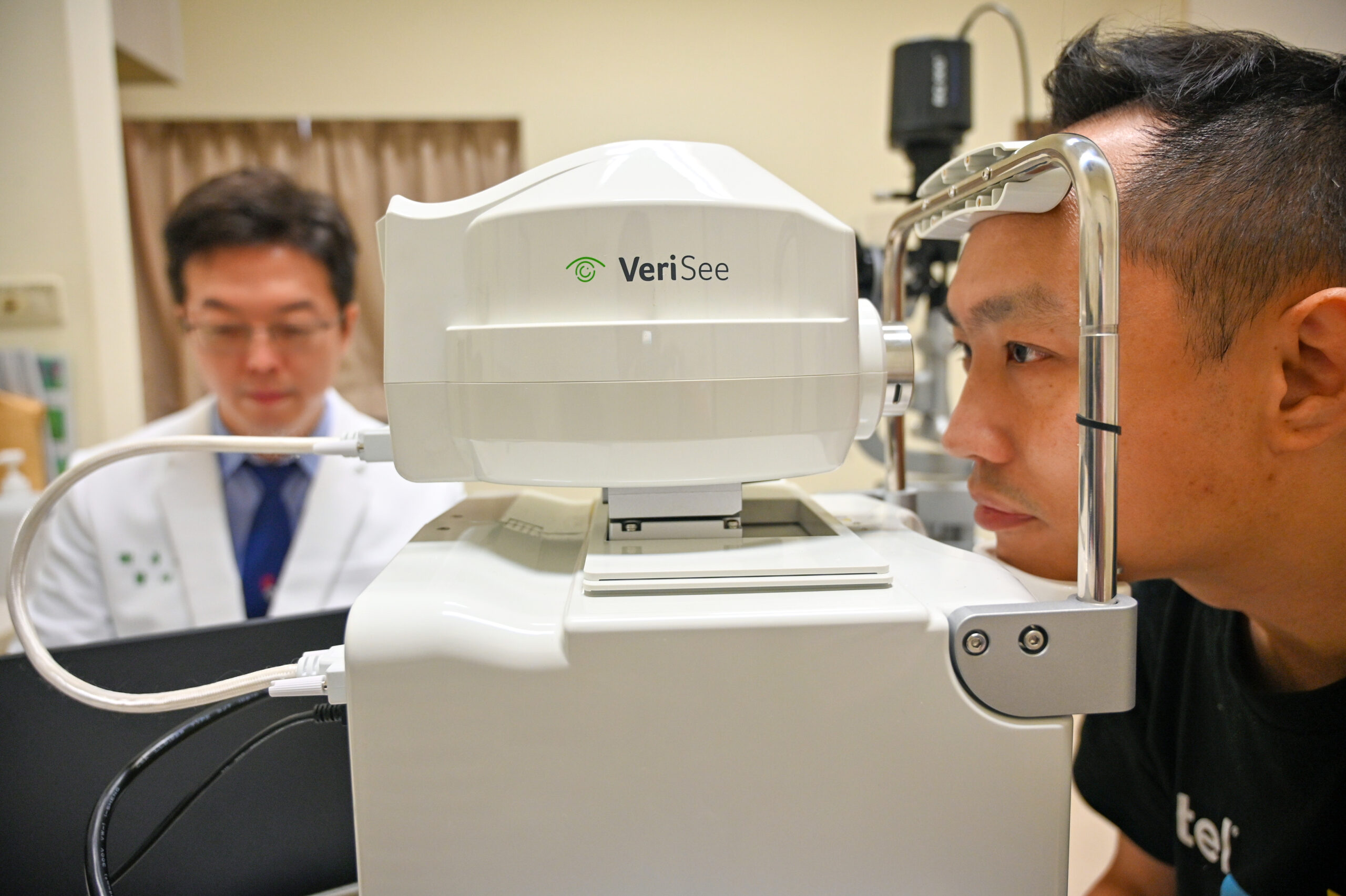
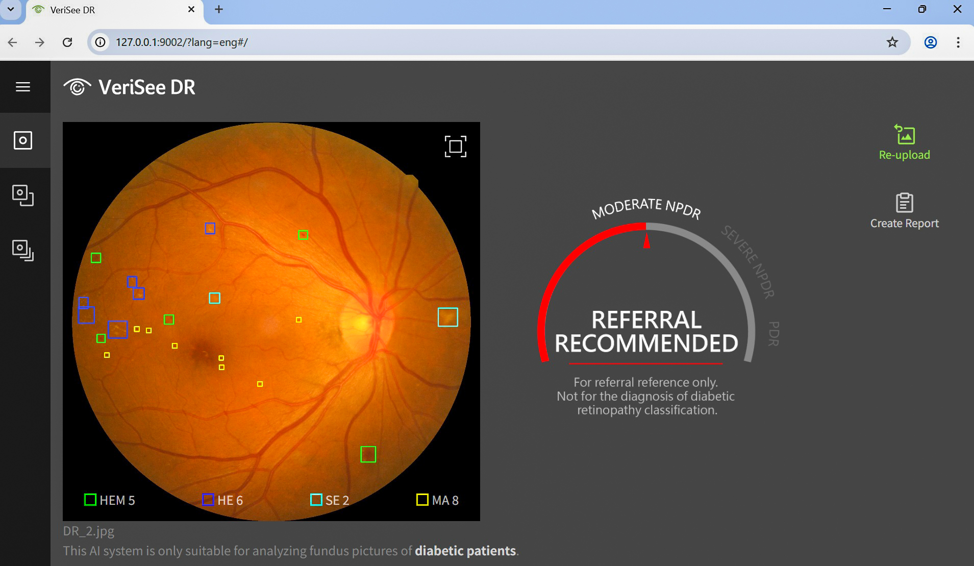
This is how the VeriSee solution works: A specialized fundus camera takes high-resolution images of the inner eye without the need to dilate pupils and sends detailed scans to a connected Acer AI PC. Specialized AI software then analyzes each image and automatically brackets eye complications in just seconds.
It also ranks potential lesion complications from moderate to severe.
That VeriSee AMD analysis today is about 95% accurate, but that number is improving as more anonymized data from other eye exams is added to train and improve the AI models.
Thanks to the AI solution’s ability to quickly detect potential eye lesions, patients no longer need to apply eye drops and wait hours for the medication to take effect prior to an eye exam in a packed clinic.
And the portability of the device – the camera and AI PC can be transported in a luggage-size case – and its ability to deliver results without an internet connection mean it can be used to examine patients in rural areas, far from large hospitals.
Hsieh stresses that the AI-backed solution doesn’t replace actual doctors. Instead, it’s a valuable tool that boosts the standard of patient care.
“The information provided by AI is a reference – a snapshot that aids our diagnosis. Although that reference is reliable, we still need medical professionals to analyze and convey the results to patients that will help them understand what the next steps and options are,” explained Hsieh.
It’s also a handy aid in teaching primary care doctors of diabetic patients and the elderly, who are mainly general practitioners and internal physicians, not eye specialists.
“A key benefit in using VeriSee DR and VeriSee AMD is the software makes it easy for us to teach other doctors who are not familiar with these lesions in the eye fundus. It saves us time as the lesions are automatically marked, making it a useful tool when we coach and train new doctors on how to make the diagnosis,” Hsieh said.
Intel-Based Hardware and Software Work in Tandem
This solution is available today from Acer Medical in Taiwan (a subsidiary of Acer Group), a specialist in advancing AI-driven medical imaging by bringing AI inferencing to the people and places on the front lines, where it’s needed most. That includes medical institutions like National Taiwan University Hospital, a national teaching medical center staffed by 7,500 medical experts.
In addition to VeriSee DR and VeriSee AMD, Acer Medical also provides hospitals with a third AI-backed screening software: VeriOsteo OP.
It’s approved for use by the Taiwan Food and Drug Administration and uses X-ray images to detect osteoporosis, a common bone mineral density (BMD) abnormality.
By analyzing chest X-ray images, VeriOsteo OP predicts the BMD and provides a recommendation. This information assists healthcare professionals in assessing the risk of BMD abnormalities. Early detection is crucial before the disease progresses to critical stages.
All three solutions are powered by Intel-backed hardware (Intel Core Ultra processors) and software (OpenVINO™ tool kits) working in tandem with Acer Medical’s proprietary algorithms.
OpenVINO Cuts Startup Time, Quickens Analysis
“With OpenVINO, we have reduced the software’s startup time by 70%, cut analysis time per image by 90%, reduced memory usage by 90%,” said Dr. Allen Lien, chairman and CEO of Acer Medical.
“These AI solutions provide rapid screening for ophthalmic diseases and abnormal bone mineral density, assist hospitals and doctors to improve the accuracy and speed of screening, and allow physicians to intervene and treat diseases early.”
Early intervention is key, stressed Lien.
Macular degeneration cases can lead to total blindness if the disease isn’t detected and treated early. With VeriSee DR’s AI-assisted diagnostic software able to detect lesions with up to 93% accuracy, treatment can begin quickly.
With VeriOsteo OP, doctors can detect bone abnormalities by analyzing a simple X-ray.
“When we apply AI-tuned algorithms to the analysis of chest X-rays, the algorithm will scan images, flag problematic areas and alert doctors to the presence of osteoporosis,” said Lien, explaining the advantages VeriOsteo OP can provide versus the current diagnosis method, which utilizes specialized bone-scanning machines.
Scaling Solutions and Adding Countries of Service
All three Acer Medical solutions are available today in more than 13 countries including Taiwan, Thailand, Malaysia and Indonesia. That list will soon expand as clinical trials are completed in more countries.
In Hsieh’s National Taiwan University Hospital in the heart of Taipei, plans are underway to scale these solutions to more clinics on campus and expand their use across more branches of medicine.
“We are currently using these AI-backed solutions in our Ophthalmology Department, along with the departments of Endocrinology and Family Medicine. In the future, we’re looking forward to making this available for senior medical care,” Hsieh said.
And, as more AI-backed medical tools become available to doctors everywhere, Hsieh remains confident AI is here to help.
“I really enjoy seeing the progress of science, especially in the field of artificial intelligence, because I think AI is just not a fancy tool. It’s really something that can help us, not only in the medical field, but I think in all fields across society,” Hsieh said.
“We have witnessed so many things that have been touched and changed by AI. It’s not something to fear, but rather, a valuable companion that can help us change the world for the better.”